Basic notions about the light microscope
Everything we need to know about the microscope and microscopy.
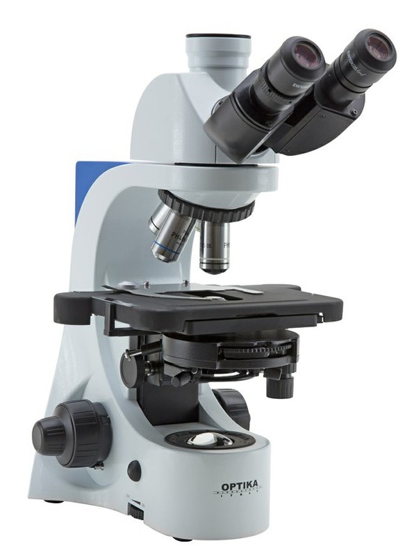
The microscope is a complex optical instrument, made up of mechanical and optical parts or lenses, and whose main objective is to be able to visualize microorganisms and structures not visible to the naked eye. Anton Van Leeuwenhoeck (1632-1723) is the father of modern microscopy, developed the light microscope that we know today, and was the first scientist to observe bacteria, protozoa, sperm, and cell tissues.

In an optical microscope we distinguish different parts, which can be classified according to are parts or optical system and parts or mechanical system.
The optical system:
He is in charge of forming and increasing the image that we want to observe. The lens and tube system captures and directs the light beam to the human eye.
Human eye: The outgoing light beam from the microscope is focused on the lens and retina of the eye.
Eyepiece: Lens that captures and enlarges the image formed on the lenses. It is the part of the microscope closest to the human eye. There are monocular, binocular, and trinocular microscopes.
Objective (s): Lens located on the revolver, captures the image on the slide and enlarges the image according to the magnification of each objective (10.40, 60, 100x) and then transmits it to the eyepiece.
Condenser: Lens that condenses light rays and projects them onto the preparation.
Diaphragm: Regulates the amount (intensity) of light entering the condenser.
Focus: Directs light rays towards the condenser.
The mechanical system:
It is the one that gives support, movement and focus to the sample that we want to observe.
Tube: It is the camera obscura that carries the eyepiece and objectives. Can be attached to the arm to allow focus
Revolver: It is the piece that carries the different objectives. It has a rotary movement to be able to choose what objective and magnification we want for observation.
Macro metric screw: Moves the stage up and down to zoom the sample in or out of the target to focus the preparation.
Micro metric screw: It is the fine focus system of the microscope, it serves to precisely focus the preparation.
Stage: It is the horizontal platform where we place the preparation. It usually has tweezers to hold the sample. The holding of the sample can be fixed or mobile in the X and Y axis (mechanical stage).
Arm: It is the structure that holds the tube, the stage and the focus screws.
Foot: It is the lower part of the microscope, which serves as a support surface. It gives us stability.
Image of the parts of an optical microscope (click to view)

The total or maximum magnification of a microscope will be obtained by multiplying the magnification of the objective by the magnification of the eyepiece, for example:
40X Objective x 10X Eyepiece = 400X
The increase in objectives is variable and we can find objectives of:
4X, 10X, 20X, 40X, 60X and 100X
The magnification of the eyepiece is always more limited and the most common are WF eyepieces of between 10 and 15X.
To observe a standard sample or fine cut it will be worth it with objectives between 4 and 40X. With these increases we will be able to see cut sections of leaves, stems, tissues and cellular structures such as protozoa, amoebae, bacteria, pollen, etc.
The 60 to 100X objectives are high magnification objectives for cellular observation in more detail, in these cases the working distance is greatly reduced and it is necessary to use immersion oil to increase the resolving power and not damage the lens optics. We will use these increases if we want to observe cells in detail inside them (cellular structure).
Another optical characteristic to take into account will be the power of resolution or the ability to distinguish two points or very close objects. In microscopy there is a minimum resolution capacity of 0.2 micrometers.
Finally, the correction power of the optical aberrations that appear in the observation is elementary. There are several types of lenses depending on your optical treatment:
Achromatic: With correction in the green and yellow field.
Platochromatic: Achromatic with good correction of the visual field curvature, the edges appear focused.
Apochromats: Correct all color errors
Apochromatic planes: Correct color and curvature errors
Parts and characteristics of a target (click to view)
Example of section or cut of a corn stalk (click to view)
There are many varieties of microscopes depending on their use. Beyond the simple optical microscope, technology during the XX and XXI centuries has developed an infinite number of applications to observe different types of materials, cells and tissues. Microscopy has and has played a fundamental role in the advancement of biomedicine and in this field in particular the greatest advances in techniques such as fluorescence and phase contrast have been developed.
Finally, the development of electron microscopes from 1937 and now digital microscopes have completed and sophisticated the world of cell observation.
Currently we find all these types of microscopes:
- Optical microscope
- Simple microscope
- Inverted microscope
- Ultraviolet light microscope
- Fluorescence microscope
- Petrographic microscope
- Darkfield microscope
- Phase contrast microscope
- Polarized light microscope
- Confocal microscope
- Electronic microscope
- Transmission electron microscope
- Scanning electron microscope
- Field ion microscope
- Scanning probe microscope
- Tunnel effect microscope
- Atomic force microscope
- Virtual microscope
Pollen image obtained by a scanning electron microscope (click to view)

A last type of microscope to highlight and of special use are the stereomicroscopes or binocular loupes. This instrument, at a lower magnification than a biological microscope (10-40X), can be binocular or trinocular (adapt a camera) and its use is focused on the observation of larger living or dead elements. With it, insects, fungi, plants, mosses, lichens, etc. are observed and identified, as well as being used to analyze industrial, construction or electronic materials.
Other related articles
 March 16, 2020
Raig in the face of the Covid-19 Coronavirus crisis
How does Raig deal with this crisis? What does it mean for …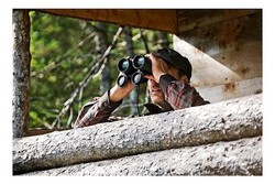 Feb. 29, 2020
The weight of the binoculars, a key factor.
The weight of the binoculars is a primary factor in choosing a …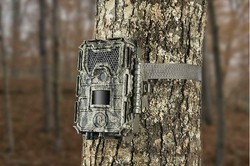 Nov. 2, 2019
Photo-trapping cameras
What is a phototrapping camera and what is it used for? What … Oct. 26, 2019
Problems and errors in rain measurement
In torrential weather episodes, we often find differences between analog and manual … Oct. 5, 2019
Opinions of our clients
Receive our news